

Tendons heal slowly in nearly everyone because the load-bearing core of an adult tendon is laid down during childhood growth and is essentially never rebuilt: carbon-14 dating of 28 human Achilles tendons found the core carried the atmospheric carbon signature of the donor's first roughly 17 years of life, while muscle from the same bodies showed continuous replacement [[1]]. The variant people quote here, COL5A1 rs12722, is associated with how likely you are to injure a ligament, and the newest meta-analysis in athletes did not confirm even that [[5]]. No published study in any species has tested it, or any other connective tissue variant, against BPC-157, TB-500 or any other peptide [[7]].
Your tendon heals slowly because its load-bearing core stopped being rebuilt when you were around 17, and we know that because of nuclear weapons testing. Bomb tests between 1955 and 1963 spiked atmospheric carbon-14, and every tissue built while levels were high carries that date stamp. When Danish researchers measured 28 human Achilles tendons, the load-bearing core matched the atmosphere of the donor's first roughly 17 years of life, while muscle from the same bodies matched recent air 1. What separates two people at month six is mostly which tissue was hurt, how much blood reaches it, age, how long the tissue had been quietly degrading before it started hurting, and what loading was done afterwards. A connective tissue gene variant is a real but small entry on that list, and it speaks to how you got injured rather than how fast you mend. (New to any of this? Start with <a href="/learn/peptides-for-beginners">peptides for beginners</a>.)
Why do tendons heal slowly?
Two things make tendon slow. It is poorly supplied with blood compared with the muscle it attaches to, so repair cells and oxygen arrive in smaller numbers. And the collagen at the centre of the tendon, the part that carries load, is not routinely replaced once you are grown.
The method behind that second point is worth a paragraph because it is unusually direct. Katja Heinemeier's group at Bispebjerg Hospital measured carbon-14 in 28 forensic samples of Achilles tendon core and 4 skeletal muscle samples, from donors born between 1945 and 1983, and compared each sample against the known atmospheric record 1. The muscle matched recent atmosphere. The tendon core did not.
The tendon concentrations of 14C approximately reflected the atmospheric levels present during the first 17 yr of life, indicating that the tendon core is formed during height growth and is essentially not renewed thereafter.Heinemeier KM, Schjerling P, Heinemeier J, Magnusson SP, Kjaer M. FASEB Journal, 2013 [[1]]
Your tendon was in trouble before it started hurting
The same group ran the method again on tendons that hurt. They compared 25 tendinopathic Achilles tendons against 10 healthy ones and found the opposite of the healthy pattern: substantial collagen renewal had occurred in the painful tendons. Modelling the carbon-14 data suggested that half the collagen in the damaged tissue had been slowly turning over for years before the person presented with symptoms 2.
That reframes the calendar. If you date your problem from the day it started hurting, you are dating it from somewhere in the middle. The person whose strain cleared in three weeks may have injured healthy tissue, while yours may have failed after a long run of abnormal remodelling that nobody could see. Two people at month six are frequently not at the same point in the same process.
Is slow tendon healing genetic?
Partly, and mostly in a way that predates the injury. The connective tissue variants that appear in consumer genetics reports were studied by comparing injured athletes against uninjured ones. That design answers one question, which is who tears something. It cannot answer how fast the torn thing repairs, because nobody in those studies measured repair.
What the collagen variant people quote does
COL5A1 is the gene for the alpha-1 chain of type V collagen, which regulates how collagen fibres assemble in tendons and ligaments. Think of it as the template that sets fibre thickness and spacing rather than the rope itself. The marker people look up, rs12722, sits in the 3' untranslated region of the gene, the tail of the RNA message that helps decide how long the message survives before the cell breaks it down. Yashna Abrahams and colleagues in Malcolm Collins's group at the University of Cape Town genotyped a different set of variants in that region (rs71746744, rs16399, rs1134170) plus a MIR608 marker in 160 people with Achilles tendinopathy against 342 controls, and showed those variants change the predicted folded shape of the message. They proposed that the shape governs message stability and therefore how much type V collagen the cell produces 3. Note that rs12722, the marker consumer reports flag, was not one of the variants they modelled. Put plainly: the mechanism is a plausible story about RNA folding that nobody has confirmed by measuring type V collagen in a human tendon, carrier against non-carrier. Treat it as a hypothesis with a citation, not a finding.
Then the evidence on whether it matters clinically splits in two.
| Meta-analysis | What went in | Verdict on COL5A1 rs12722 | Other markers |
|---|---|---|---|
| Guo et al., Journal of Orthopaedic Surgery and Research, 2022 4 | 21 observational studies of musculoskeletal soft tissue injury, searched to August 2021 | Associated with higher injury susceptibility. On stratified analysis the association sat mainly in ligament injury and in people of European descent | rs71746744 and rs3196378 trended toward higher risk; rs13946 was associated with tendon and ligament injuries on subgroup analysis; rs11103544 showed no association |
| Fukuyama et al., International Journal of Sports Medicine, 2025 5 | 31 articles in athletes, 1,687 injury cases, 2,227 controls, meta-analysis pooling 12 of them, searched to July 2023 | Not confirmed as a risk factor for tendon or ligament injury | COL1A1 rs1800012 and MMP3 rs679620 also not confirmed. Only VEGFA rs699947 reached significance, and in the protective direction (C versus A allele, odds ratio 0.80, 95% CI 0.65 to 0.98, meaning roughly 20% lower injury odds for C carriers, with the true figure somewhere between 2% and 35% lower) |
Both are competent reviews. They differ because Guo pooled general population studies of soft tissue injury and Fukuyama restricted the question to athletes, and an association that survives in mixed populations can vanish in a fitter, more heavily selected one. Fukuyama also pooled only 12 of its 31 articles across four markers, so its rs12722 result is an underpowered failure to confirm rather than a refutation. What a reader should take from the disagreement is the size of the effect being argued over. Two careful teams cannot agree that this variant changes injury odds at all, and neither of them measured healing speed.
Why an injury-risk marker cannot predict how you respond to a compound
Susceptibility was measured. Response never was. Stretching a susceptibility marker into a treatment prediction is a common error in consumer genetics. A marker earns the word predictive for a drug only when somebody has given the drug to carriers and non-carriers and measured the difference. That has been done for warfarin dosing and for clopidogrel. It has never been done for BPC-157, TB-500 or GHK-Cu, in a human or in an animal, against COL5A1 or against any other connective tissue gene.
What a risk genotype buys you is a reason to take tendon load management and rehab patience more seriously.
There is a further practical wrinkle. Rodent studies are where nearly all the tendon peptide data lives, and no rodent study genotypes rats at a human single-letter variant. So even the animal evidence cannot be read by genotype. If you want the ordered way to weigh evidence, legality and DNA when picking any compound, <a href="/learn/which-peptides-dna-decision-framework">our decision framework for which peptides your DNA should change</a> puts genetics at step five for exactly this reason.
What has been tested on a tendon, and in what
Here are the real protocols behind the compounds people bring to a sore tendon, reported as research rather than as instructions. Note how many rows are rats, and how the one human row involves no drug at all.
| Compound | What was tested | Dose and route in the study | Time before measurement | Result |
|---|---|---|---|---|
| BPC-157 8 | Surgically transected and repaired Achilles tendon in 32 male Sprague-Dawley rats, 8 per arm split between biomechanical testing and microscope work | 10 mcg/kg/day, injected into the abdominal cavity | 4 weeks | Higher maximum load to failure than controls but not statistically significant; tissue-damage scores numerically better without reaching significance |
| TB-500 8 | Same 32-rat Achilles study, separate arm | 60 mcg/kg/day, injected into the abdominal cavity | 4 weeks | Significantly higher maximum load to failure (p < 0.05), and lower Bonar and Movin scores (p = 0.016 and p = 0.017), the two standard microscope scales for tendon tissue damage, where lower means healthier-looking tissue |
| BPC-157 plus TB-500 8 | Same study, combination arm | Both doses together, abdominal cavity | 4 weeks | Lower Movin score than untreated controls (p = 0.040), but no additional benefit over either compound alone |
| Full-length thymosin beta-4 9 | Severe dry eye in 9 people. Not a tendon, and not the injected fragment | 0.1% eye drops, six times daily for 28 days | 56 days | 35.1% less ocular discomfort (p = 0.0141) and 59.1% less corneal staining (p = 0.0108) versus vehicle |
| BPC-157, entire human record 7 | 544 articles screened from 1993 to 2024; 36 met inclusion | Not applicable | Not applicable | 35 preclinical studies and 1 clinical study, a retrospective series in which 7 of 12 patients reported relief beyond 6 months after an injection into the knee for unspecified chronic knee pain |
| GHK-Cu | Nothing in a tendon, in any species | Not applicable | Not applicable | No tendon evidence of any kind. Its human record is topical cosmetic use |
| Heavy-load eccentric heel-drops 6 | Chronic Achilles tendinopathy in 15 recreational athletes, mean age 44.3 | Twice-daily eccentric calf training, no compound | 12 weeks | All 15 returned to pre-injury running; all 15 conventionally treated comparators were ultimately operated on |
One row deserves saying out loud rather than leaving in a table: GHK-Cu has no tendon evidence in any species. It appears on tendon stacks because it appears on stacks, and its human record is topical and cosmetic. That one is mostly marketing.
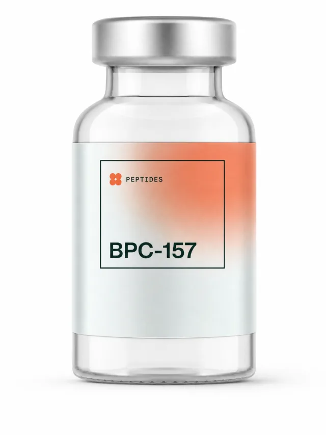
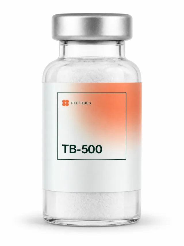
The two compounds people choose between
The combination of the two is the best-known blend in this space, sold and discussed as a named stack.
That head-to-head tested the combination as its fourth arm. It did beat untreated controls on one tissue damage score (p = 0.040), and the authors were direct about the rest of the outcome: combined BPC-157 and TB-500 treatment did not confer additional benefits compared with either agent alone 8. They suggested the two may converge on shared downstream pathways, and flagged that as a hypothesis needing confirmation rather than a finding. With 8 rats per arm split across two kinds of measurement, the honest reading is that the combination has not been shown to beat the parts, not that it has been ruled out.
What moves a stubborn tendon
How a tendon rebuilds, and where the compounds are aimed
The human evidence for that last step is worth reading in detail because it is unusually clean for a rehab study. Håkan Alfredson's group in Umeå took 15 recreational athletes, mean age 44.3, all with chronic Achilles tendinopathy and pain severe enough to stop them running, and all of whom had already failed conventional care. Twelve weeks of heavy-load eccentric calf training returned all 15 to their pre-injury running level, with calf strength on the injured side no longer significantly different from the healthy side. Eccentric here means the lowering half of the movement: raising up on both feet, then lowering the injured heel slowly below the step under body weight. A comparison group of 15 athletes with the same diagnosis went through rest, anti-inflammatories, orthoses and physiotherapy; in no case was that successful and all 15 were ultimately operated on 6.
Fifteen people is a small study and it was not randomised, so weigh it accordingly. It is still the strongest human evidence on this page by a wide margin, and it has held up as the basis of Achilles rehab for nearly three decades. If you want to try it, the protocol has a name, so ask a physiotherapist for the Alfredson protocol and have them check the tendon first. For the tendon-specific compound evidence and the protocols circulating online, see <a href="/peptides-for/tendonitis">peptides for tendonitis</a>.
Cost, storage and what is unknown
Peptides for tendon use are sold as research chemicals, labelled not for human consumption, priced per vial by milligram content, with no pharmaceutical manufacturing standard behind them. What follows describes what circulating protocols involve; it is not an endorsement of buying or using any of them, and none is approved for human use. The cost that matters is how many vials a multi-month schedule implies, because circulating tendon protocols run 8 to 12 weeks, and 16 to 24 weeks for chronic cases (see <a href="/learn/peptide-cycling-protocol">how peptide cycling protocols are structured</a>). The 2025 systematic review flags unregulated manufacturing and contamination as a source of possible adverse effects 7, which is why buyers in this market treat a batch-specific certificate of analysis as the floor when a plan depends on consistent content across months. How the main suppliers handle batch testing is broken down in <a href="/compare">our vendor comparison</a>. Disclosure: some outbound links on PeptidesDNA are affiliate links and we may earn a fee at no extra cost to you; this never affects what we list or how we rate it.
None of what follows is a recommendation to self-administer. Storage is the step most often reported wrong: these compounds ship as a freeze-dried powder, which is stable refrigerated, and once mixed with bacteriostatic water they become a solution with a much shorter usable life that needs to stay cold. The mechanics are documented in <a href="/learn/how-to-reconstitute-peptides">how to reconstitute peptides</a> and <a href="/learn/peptide-injection-hygiene-guide">peptide injection hygiene</a>, which describe what people report doing rather than what you should do. The commonly circulated figures for tendon use, 250 to 500 mcg of BPC-157 daily and 2.5 to 5 mg of TB-500 twice weekly, are conventions from online protocols rather than trial-derived doses; there is no established human dose for either compound, the 2025 systematic review found no clinical safety data at any dose 7, and the rat studies used a different route entirely. The figures themselves and where they came from are traced in <a href="/learn/bpc-157-dosage-guide">the BPC-157 dosage guide</a>.
What your DNA can add here
Knowing you carry a connective tissue risk variant is a reason to be stricter about load management, warm-ups and rehab patience. It is not a reason to buy any particular compound. That is the whole actionable content of the finding, and anyone selling you more than that is going beyond the published data.
If you want to check your own genotype, rs12722 is carried on most consumer arrays and appears in a 23andMe or AncestryDNA raw file as a two-letter code, though strand orientation can make the letters differ from the published C and T naming. What a raw file can and cannot resolve, including why a flagged variant is a starting point for a conversation rather than a result, is covered in <a href="/learn/23andme-peptide-guide">what your 23andMe raw data can tell you about peptides</a>. The same limits apply to the joint and tendon markers people ask about on <a href="/peptides-for/joint-pain">peptides for joint pain</a>, and to the general ranking of compounds in <a href="/learn/best-peptides-for-healing">the best peptides for healing</a>.
The reason to look at genetics for tendons is expectation-setting. Knowing your tendon core was mostly built before you were 18, and that the tissue may have been remodelling abnormally for years before it hurt, is more useful for planning a recovery than any single-letter variant currently on the market. How we build and grade our marker panel, and where we say the evidence runs out, is set out in <a href="/methodology">our methodology</a>.
Link · Heinemeier et al., FASEB Journal, 2013Lack of tissue renewal in human adult Achilles tendon is revealed by nuclear bomb 14CThe bomb-pulse carbon dating study behind the 17-year figure. 28 Achilles tendon core samples and 4 muscle samples, donors born 1945 to 1983.pubmed.ncbi.nlm.nih.govUpload your raw DNA data and we will report your genotype at every marker on the PeptidesDNA panel, with the evidence level stated for each one, including the many, like the tendon markers on this page, where the answer is that no study has tested it. The report is educational, does not predict how you will respond to any compound, and is not a recommendation to use one.
Get your DNA report- Susceptibility was measured, response never was
Every connective tissue variant quoted for tendons comes from studies that compared injured people against uninjured people. Those studies recorded who got hurt. None of them gave anyone a compound, so none can say how a carrier responds to BPC-157, TB-500 or anything else, and no published study in any species has tried. What the carbon-14 work adds is why the question is aimed at the wrong target in the first place: the load-bearing core of an adult Achilles carries the atmospheric carbon signature of the donor's first roughly 17 years of life, and tendinopathic tendons had been quietly remodelling for years before they hurt. Slow tendon healing is normal biology for everyone long before it is anyone's genetic fault.
Sources9
- Heinemeier KM, Schjerling P, Heinemeier J, Magnusson SP, Kjaer M. Lack of tissue renewal in human adult Achilles tendon is revealed by nuclear bomb 14C. FASEB Journal, 2013;27(5):2074-9.
- Heinemeier KM, Schjerling P, Øhlenschlæger TF, Eismark C, Olsen J, Kjær M. Carbon-14 bomb pulse dating shows that tendinopathy is preceded by years of abnormally high collagen turnover. FASEB Journal, 2018;32(9):4763-4775.
- Abrahams Y, Laguette MJ, Prince S, Collins M. Polymorphisms within the COL5A1 3'-UTR that alters mRNA structure and the MIR608 gene are associated with Achilles tendinopathy. Annals of Human Genetics, 2013;77(3):204-14.
- Guo R, Ji Z, Gao S, et al. Association of COL5A1 gene polymorphisms and musculoskeletal soft tissue injuries: a meta-analysis based on 21 observational studies. Journal of Orthopaedic Surgery and Research, 2022;17(1):129.
- Fukuyama Y, Murakami H, Iemitsu M. Single nucleotide polymorphisms and tendon/ligament injuries in athletes: a systematic review and meta-analysis. International Journal of Sports Medicine, 2025;46(1):3-21.
- Alfredson H, Pietilä T, Jonsson P, Lorentzon R. Heavy-load eccentric calf muscle training for the treatment of chronic Achilles tendinosis. American Journal of Sports Medicine, 1998;26(3):360-6.
- Vasireddi N, Hahamyan H, Salata MJ, Karns M, Calcei JG, Voos JE, Apostolakos JM. Emerging use of BPC-157 in orthopaedic sports medicine: a systematic review. HSS Journal, 2025;21(4):485-495.
- Biçer O, Adanir O, Güleryüz Y, Balci EC, Dinçel YM, Yenigün MY, Aydin C, Bayrak BY. Effects of BPC-157 and TB-500 on Achilles tendon healing in rats: a histopathological and biomechanical study. Joint Diseases and Related Surgery, 2026;37(3):822-837.
- Sosne G, Dunn SP, Kim C. Thymosin β4 significantly improves signs and symptoms of severe dry eye in a phase 2 randomized trial. Cornea, 2015;34(5):491-6.
This article is for informational and educational purposes only. It is not medical advice and does not diagnose, treat, cure, or prevent any disease. Consult a qualified healthcare professional before starting any peptide protocol. Individual results vary. Some outbound links are affiliate links, at no extra cost to you.

Frequently asked questions
Why does my tendon take so long to heal?
Mostly because tendon is slow tissue in everyone. Carbon-14 dating of 28 human Achilles tendons found the load-bearing core carries the atmospheric carbon signature of the donor's first roughly 17 years of life, meaning it is built during height growth and essentially not renewed afterwards, while muscle from the same donors turned over continuously. Tendon is also poorly supplied with blood compared with the muscle it attaches to, so repair cells arrive in smaller numbers. On top of that, tendons with long-running degeneration show years of abnormal collagen turnover before symptoms appear, so the injury you are counting weeks from may have been developing long before it hurt.
Is slow tendon healing genetic?
Partly, and in a narrower way than most consumer genetics copy suggests. Connective tissue variants such as COL5A1 rs12722 were studied by comparing injured athletes against uninjured ones, which measures who gets hurt rather than who repairs quickly. Even for injury risk the evidence is contested: a 2022 meta-analysis of 21 observational studies found an association concentrated in ligament injury and in people of European descent, while a 2025 meta-analysis restricted to athletes (31 articles, 1,687 cases, 2,227 controls) did not confirm it. Healing speed itself has never been the outcome in any of these studies.
Does COL5A1 tell me which peptide to use for a tendon?
No. No published study in any species has tested COL5A1 genotype, or any other connective tissue variant, against response to BPC-157, TB-500, GHK-Cu or any other peptide. Rodent tendon studies do not genotype animals at human variants, so even the animal literature cannot be read by genotype. Any protocol that assigns a specific compound to a specific genotype for tendon repair is extrapolating from an injury-risk marker, which is a different question. The variant supports stricter load management and more patience with rehab.
Do BPC-157 or TB-500 have human tendon evidence?
Neither has a published human tendon trial. A 2025 systematic review of BPC-157 in orthopaedic sports medicine screened 544 articles from 1993 to 2024 and included 36: 35 preclinical, meaning animals and lab dishes, and 1 clinical, a retrospective series in which 7 of 12 patients reported relief beyond six months after an injection into the knee for unspecified chronic knee pain. The review also states that no clinical safety data were found. The human trial data often attributed to TB-500 belongs to full-length thymosin beta-4 delivered as an eye drop in a 9-patient dry eye trial, which is a different molecule by a different route. The only head-to-head is a 32-rat Achilles study in which TB-500 alone had the biomechanical advantage and the combination beat neither compound alone.
This article is for informational and educational purposes only. It is not medical advice and does not diagnose, treat, cure, or prevent any disease. Consult a qualified healthcare professional before starting any peptide protocol. Individual results vary. Some outbound links are affiliate links, at no extra cost to you.